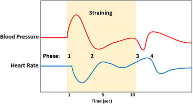
The Valsalva Maneuver is a test used to evaluate autonomic function by measuring the cardiovascular system’s response to a forced exhalation against a closed airway. This maneuver helps assess the autonomic control of heart rate and blood pressure. And also used in clinical practice in the diagnosis and treatment of SVTs and, occasionally, for the assessment of heart failure and left ventricular dysfunction Here’s a detailed step-by-step guide on how to perform and interpret the Valsalva Maneuver:
1. Preparation:
- Explain the Procedure: Explain the test to the patient, ensuring they understand the steps and purpose. This helps reduce anxiety and ensures better cooperation.
- Ensure Comfort: The patient should be comfortably seated or lying down in a quiet environment to minimize external influences.
2. Equipment Needed:
- Blood Pressure Monitor: An automatic or manual sphygmomanometer to measure blood pressure.
- Heart Rate Monitor: An ECG machine or a heart rate monitor to record heart rate.
- Manometer: To measure the pressure generated during the maneuver (optional).
3. Baseline Measurements:
- Resting Heart Rate and Blood Pressure: Record the patient’s resting heart rate and blood pressure after they have been resting quietly for at least 5 minutes.
4. Performing the Maneuver:
4.1 Instructions to the Patient:
Instruct the patient to take a deep breath and then exhale forcefully into a mouthpiece or against a closed glottis (like blowing into a syringe) for 15 seconds. The goal is to maintain a pressure of about 40 mmHg. Signs of adequacy include neck vein distension, increased tone in the abdominal wall muscles, and a flushed face. The patient should maintain the strain for 10 to 15 seconds and then release it and resume normal breathing. A modified Valsalva maneuver, which involves the standard strain (40 mmHg pressure for 15 seconds in the semirecumbent position) followed by supine repositioning with 15 seconds of passive leg raise at a 45 degree angle, has been shown to be more successful in restoring sinus rhythm for patients with SVT .
4.2 Monitoring:
During this period, continuously monitor and record the heart rate and blood pressure. Ideally, the continuous monitoring is performed with 12-lead electrocardiography, yielding the most information about the heart rhythm and tachycardia, but if this is not available or practical then continuous single-lead telemetry monitoring should be performed. Patients who are performing the Valsalva maneuver for diagnostic purposes in this setting should have continuous blood pressure monitoring along with continuous heart rate monitoring (single-lead telemetry is adequate here) during the maneuver. When noninvasively monitoring blood pressure responses using a blood pressure cuff, the cuff should be inflated to approximately 15 mmHg above the patient’s resting systolic blood pressure, and the examiner should auscultate the brachial artery throughout the maneuver and for 15 to 30 seconds afterward.
4.3 Phases of the Valsalva Maneuver:
Phases 1 and 2 occur during the active strain phase of the Valsalva maneuver, while phases 3 and 4 occur after the strain phase has been completed.
4.3.1 Phase I (Initial Strain):
A transient rise in blood pressure (by a >15 mmHg) as intrathoracic pressure increases.
4.3.2 Phase II (Continued Strain):
A decrease in venous return and cardiac output, leading to a drop in blood pressure (return of the systolic blood pressure to baseline below the 15 mmHg increase) and a compensatory increase in heart rate.
4.3.3 Phase III (Release of Strain):
An immediate drop in blood pressure as the strain is released. abrupt fall in systolic blood pressure below baseline. Phase 3 occurs due to an acute decrease in intrathoracic pressure.
4.3.4 Phase IV (Recovery):
Is identified by a secondary rise in systolic blood pressure >15 mmHg above baseline. Phase 4 occurs because of a reflex sympathetic response to the decrease in systolic blood pressure encountered during phase 3. Relative bradycardia may occur during this phase. Recovery and Post-Test Measurements:
- Resting Phase: Allow the patient to rest quietly for 1-2 minutes after the maneuver.
- Record Measurements: Record the heart rate and blood pressure during the recovery phase.
4.4 Interpretation of Results
4.4.1 Normal Response:
- Heart Rate: A marked increase during Phase II and a decrease during Phase IV.
- Blood Pressure: An initial rise in Phase I, a drop during Phase II, a further drop in Phase III, and an overshoot in Phase IV before normalizing.

4.4.2 Abnormal Response:
- Blunted Heart Rate Response: May indicate autonomic dysfunction.
- Absent overshoot: May suggest impaired autonomic control or cardiovascular dysfunction (moderately decreased left ventricular ejection fraction).
- The “square wave” response is characterized by the presence of Korotkoff sounds during the entire straining phase (indicating a sustained rise in blood pressure following phase 1) and an absence of the expected rise in systolic blood pressure during phase 4. This response is associated with a severely decreased ejection fraction.
- Patients taking beta-blockers typically show a blunted blood pressure response to the Valsalva maneuver.
4.5 Example Values
4.5.1 Normal Valsalva Ratio:
The ratio of the longest RR interval after the maneuver to the shortest RR interval during the maneuver should be greater than 1.5.
4.5.2 Blood Pressure Response:
- Phase I: Increase in systolic blood pressure.
- Phase II: Decrease in systolic blood pressure.
- Phase III: Transient decrease in systolic blood pressure.
- Phase IV: Overshoot of systolic blood pressure above baseline.
5. Practical Tips
- Patient Cooperation: Ensure the patient understands and cooperates fully during the maneuver.
- Consistent Pressure: Encourage the patient to maintain a consistent pressure of 40 mmHg if using a manometer.
- Monitoring: Continuous monitoring is crucial to accurately capture the rapid changes in heart rate and blood pressure.
6. Safety and Precautions
- Supervision: Perform the test under medical supervision, especially in patients with known cardiovascular conditions.
- Contraindications: Avoid the maneuver in patients with recent myocardial infarction, severe coronary artery disease, or other high-risk cardiovascular conditions.
7. Heart rate responses for patients with SVT
For a patient with SVT in whom the Valsalva maneuver is performed for both diagnostic and/or therapeutic purposes, the potential heart rate and rhythm responses are similar to those that can be seen following any vagal maneuver or the administration of adenosine. Potential outcomes include slowing of sinoatrial nodal activity, block at the AV node “unmasking” atrial activity, termination of the SVT, or no response (generally indicating inadequate performance of the technique).
