In persons with diabetes, the diagnostic accuracy of clinical examination for the presence of peripheral arterial disease (PAD) is low. Therefore, in any person with a foot ulcer, objective assessment of the perfusion in the foot is warranted with tests described below. These test are also advised when PAD is suspected in a person without a foot ulcer. Â
Materials required
- Hand-held 5-10 mHz Doppler device.
- Transducer gel.
- Select blood pressure cuff of sufficient size to be placed around the upper arms and calves (approx. 40% extra to wrap around).
Â
Measurement conditions
- Quiet surroundings in room with comfortable temperature for the patient, such as 22âÂÂ24 °C.
- Alcohol, exercise and caffeine should be avoided for 2 hours prior to testing.
- Patient in supine horizontal position, for 10 minutes prior to the measurement.
- Both arms and lower legs should be bare.
- No tight sleeves of shirts and trousers.
- Always use the same sequence of measurements as described below.
Brachial & ankle pressures and Doppler waveforms
Brachial pressure
- Place cuff around the upper arm.
- Apply the gel over the area of the brachial artery (can be palpated first). Ensure that a clear audible signal is detected.
- Inflate the cuff to supra-systolic values, i.e. about 30 mmHg above the pressure when the signal disappears completely.
- Slowly deflate the cuff at a rate of 2âÂÂ3mmHg per second until an audible signal re-appears, the cuff pressure at that moment equals the systolic pressure in the artery.
- Record the result.
- Repeat this procedure in the other arm.
Ankle pressure and assessment of the Doppler waveform
- Place the calf cuff approximately 2 cm above the malleolus, with the tubes pointing upwards
- Apply the gel in the areas of the dorsalis pedis and posterior tibial arteries (see figure below)
- Place the Doppler probe with an angle of 40-60° pointing upstream in the area of each artery
- Slowly move the probe to select the area with the best signal.
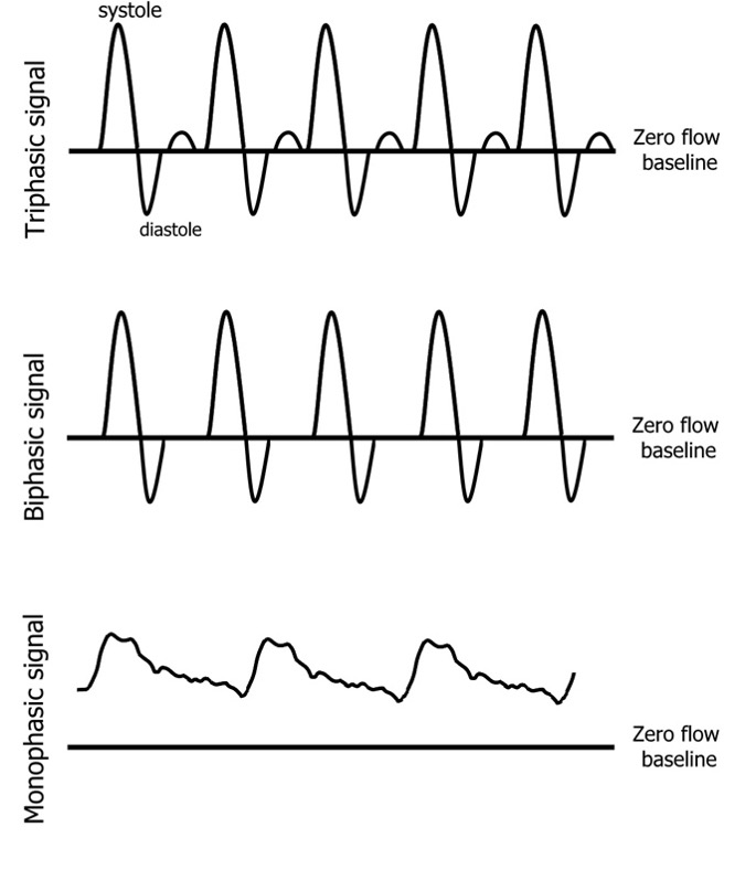
- Ideally print/ review waveform on screen of doppler machine. If the waveform is not displayed by the machine used, audibly assess the Doppler waveform and sound.
- An absent signal or a monophasic signal is abnormal and is indicative of the presence of peripheral artery disease .
- Inflate the cuff to 30 mmHg above the pressure where the pulsatile sound is lost/the visual waveform disappears.
- Slowly deflate the cuff at a rate of 2âÂÂ3mmHg per second, the systolic pressure should be taken as soon an audible waveform returns or there is a small regular upstroke of a visual waveform (which occurs before the full waveform returns). Record the result.
- After a minute rest, perform the measurement on the other artery of the same foot or if the signal was lost during the first measurement (do not reinflate the cuff during the procedure)
- Repeat these measurements on the other leg.
Calculation of ABI in persons with diabetes
To diagnose peripheral artery disease, calculate the ankle-brachial index (ABI) for each limb ABI = lower value of the dorsalis pedis or posterior tibial pressures / highest brachial pressures  by dividing the lower value of the dorsalis pedis or posterior tibial pressures of that foot by the highest of the left or right brachial pressures. This is particularly in those people with diabetes who have below knee arterial disease, which may affect only one of the tibial arteries.  The ABI has traditionally been calculated using the higher of the dorsalis pedis or posterior tibial pressures. This gives a best-case scenario of blood flow to the foot.  An ABI above 1.3 or below 0.9 is abnormal, i.e. indicative of PAD. Â
Â

